Visual Dysfunction in Parkinson’s Disease May Predict Cognitive Performance
Higher order visual tests could help stratify patients in clinical trials
05/08/2023
Paul Smyth, MD, Contributing Writer, BreakingMED™
Kevin Rodowicz, DO, Assistant Professor, St. Luke’s University/Temple University
Higher order vision function—a composite measure of skew tolerance and biological motion detection—bested retinal thickness for predicting cognitive performance in Parkinson’s disease, a prospective cohort study found.
The findings support mechanistic models for Parkinson’s dementia progression with onset in cortical structures, and shows potential for visual tests to stratify patients in clinical trials, the researchers suggested.
Higher order vision function — a composite measure of skew tolerance and biological motion detection — bested retinal thickness for predicting cognitive performance among people with Parkinson’s disease (PD), according to a prospective cohort study.
"This study confirms that higher order visual tests could be useful to stratify patients to enrich clinical trials and as predictive measures in the clinic; but that structural retinal measures are less likely to be effective in this way," the researchers added.
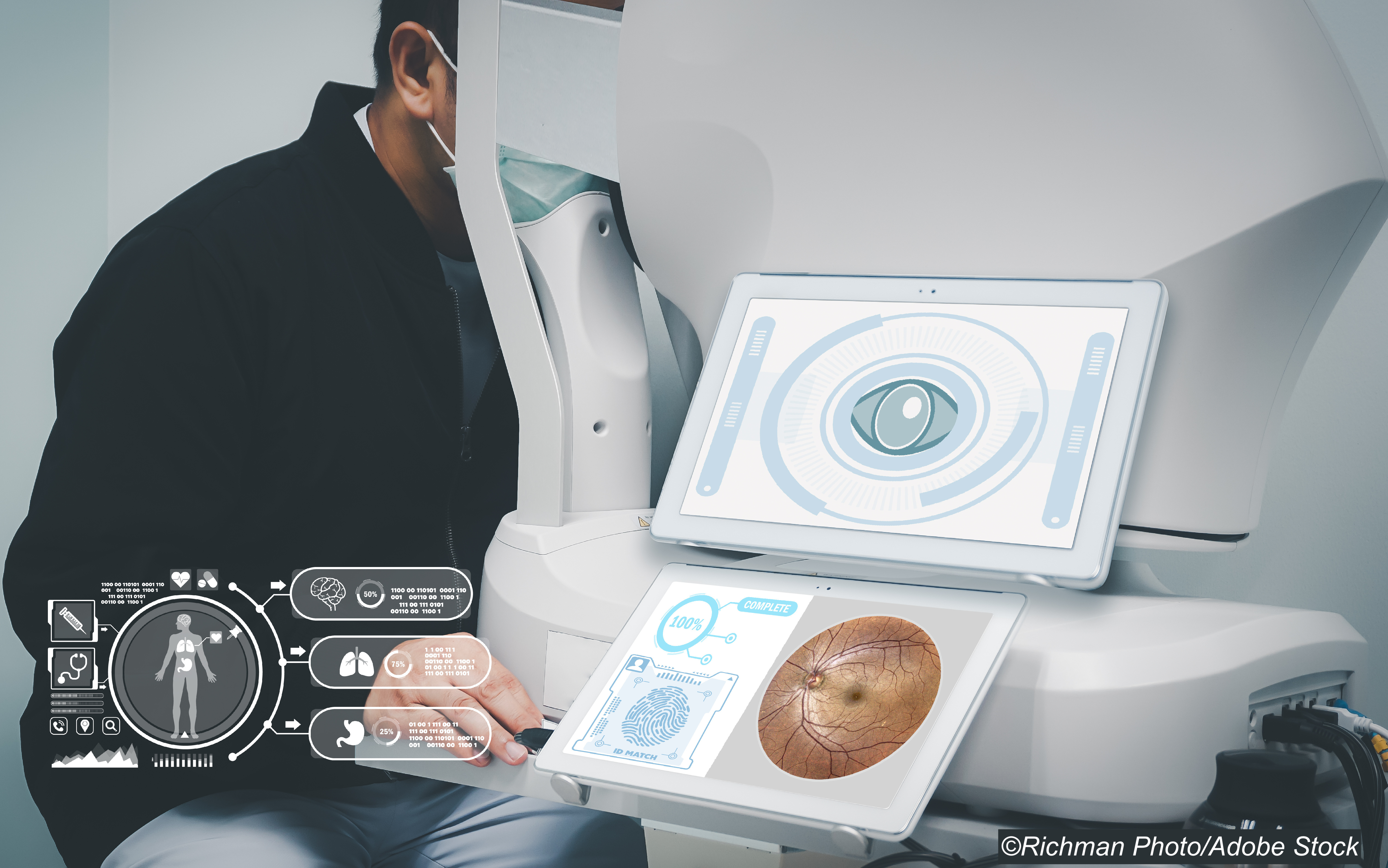
Both retinal structure — with optical coherence tomography (OCT)-defined thinning in the ganglion cell-inner plexiform layer — and higher-level visual function including visual rotation, line orientation, and spatial navigation, are affected in PD.
For 100 people with PD (mean age 64.1, 45% women) and 29 age- and sex-matched unaffected controls (mean age 64.2, 55% women), Weil and colleagues found a decline in a cognitive composite score of -0.039 units per year in the PD group.
Overall, 37 PD patients were classified as low-vision performers and 63 were classified as high-vision performers.
- For a 1-unit decrease in the baseline higher order visual score, cognitive score was predicted to decrease by 0.178 (β=0.178, P=0.0005) and the decrease per year in cognitive score would be 0.024 points greater (β=0.024, P=0.013).
- In contrast, baseline retinal thickness of the ganglion cell-inner plexiform layer did not predict cognitive scores (β=−0.013, P=0.87) or their change over time (β=0.024, P=0.12).
- Time-to-event analysis found that participants with low visual performance had increased rates of dementia (χ² (1)=5.2, P=0.022) and of a composite dementia, death, or frailty (χ² (1)=10.0, P=0.002) compared with those who had high visual performance.
"Using linear mixed-effect models adjusted for age, we showed that higher order visual dysfunction significantly predicted longitudinal cognitive decline, but that retinal thickness showed no predictive effects for cognition," Weil and colleagues wrote. "Consistent with these findings, our survival analyses showed a greater conversion to dementia, death, or frailty in patients with poorer baseline higher order visual function, but not for those with thinner retinas."
"Importantly, our mixed model analysis allowed us to specifically take into account baseline cognition, showing that these tests provide additional information to standard cognitive tasks and confirm the utility of these or similar higher-order visual tests as stratification tools for the clinic or in clinical trials," they emphasized.
The researchers included participants who met a clinical diagnosis of PD per Queen Square Brain Bank Criteria, and who were recruited from U.K. clinics between October 2017 and November 2018. All were within 10 years of diagnosis. Those with a diagnosis of dementia, a Mini-Mental State Examination (MMSE) score of <25 (maximum score 30, lower scores indicate worse cognitive performance), or ophthalmic disease sufficient to impair visual acuity were excluded.
Participants had clinical and ophthalmic assessments at baseline, and clinical assessments at 18 and 36 months.
General cognitive function was assessed with the MMSE and Montreal Cognitive Assessment (MoCA). MoCA scores contributed to a composite cognitive score that also included domain-specific assessments of memory, language, visuospatial ability, executive function, and attention compared as a z-score.
OCT data included ganglion cell-inner plexiform layer, inner nerve layer, and peripapillary retinal nerve fiber layer measurements. Higher order visuoperception was measured with summed z-scores of two visual parameters that are affected in PD and have been associated with worse cognition, the authors noted: the Cats-and-Dogs test to measure visual skew and a biological motion test.
Weil and colleagues proposed that two competing theories may explain retinal and higher order visual dysfunction in PD.
"One model suggests that degenerative changes arise in cortical regions and spread to the retina through a process such as retrograde trans-synaptic axonal degeneration; the other proposes that both cortical and retinal cells are vulnerable to degeneration in PD, and that de-arborization occurs idiosyncratically throughout the nervous system, which might affect retina and cortex at similar times when examining a population," they wrote.
"Our prospective study supports the former model, with primary neurodegeneration likely affecting cortical regions before involving distal areas such as the retina," they added.
Limitations of the study include diagnoses of atypical parkinsonism or mild cognitive impairment received by eight patients during follow-up (six in the PD group and two in controls). All were excluded from further analysis. The rate of these diagnoses was in line with other published longitudinal PD cohorts, the authors noted.
Weil and colleagues also acknowledged that other retinal measures, like electroretinography, may have greater sensitivity to predict outcomes in PD than that seen with OCT in this study.
Disclosures
This work was funded by grants from UCLH Biomedical Research Centre Grant.
Weil is supported by a Wellcome Clinical Research Career Development Fellowship. This work was funded by grants from UCLH Biomedical Research Centre Grant, and has received honoraria from GE Healthcare and Britannia, as well as speaking honoraria from the Shirley Ryan Ability Lab.
Sources
Hannaway N, et al "Visual dysfunction is a better predictor than retinal thickness for dementia in Parkinson’s disease" J Neurol Neurosurg Psychiatry 2023; DOI: 10.1136/jnnp-2023-331083.